A mammogram is an X-ray image of the breast that can be used to screen for breast cancer or for diagnostic purposes, such as checking for symptoms or unusual findings in another imaging test.
During a mammogram, the breasts are compressed between two firm surfaces to spread the breast tissue. An X-ray then takes black-and-white images that are displayed on a computer screen and checks for signs of cancer.
Mammography plays a key role in breast cancer screening. They can detect breast cancer before it causes signs and symptoms. It has been shown to reduce the risk of dying from cancer.
How does a mammogram work?
Mammograms are X-ray images of your breasts designed to detect cancer and other changes in breast tissue. It can be used for screening or diagnostic purposes:
- Screening mammography: Screening mammography is used to detect breast changes that could be cancerous in people who have no symptoms. The goal is to detect cancer when it is small, and treatment is less invasive.
- Diagnostic mammography: Diagnostic mammography is used to check for suspicious breast changes, such as a new breast lump, breast pain, unusual skin appearance, nipple thickening, or nipple discharge. It is also used to evaluate unexpected findings in screening mammography. A diagnostic mammogram includes additional images.
Risks
Risks and limitations include:
- Mammography exposes you to low-dose radiation: However, the dose is very low, and for most people, the benefits of a regular mammogram outweigh the risks of this amount of radiation.
- A mammogram may lead to additional tests: A mammogram may lead to additional tests. Other tests may be needed if something unexpected is detected on the mammogram. This may include additional imaging tests such as an ultrasound and a procedure (biopsy) to remove a sample of breast tissue for the laboratory. However, most findings on mammograms are not cancer.
- Screening mammography can’t detect all cancers: Some cancers detected by a physical exam may not be seen on a mammogram. If the tumor is very small or in an area that is difficult to see on a mammogram, such as an armpit, it may be missed.
- Not all cancers detected by mammography are curable: Some breast cancers are aggressive, grow quickly, and spread quickly to other body parts.
Prepare
To prepare for a mammogram:
- Schedule the test when the breasts are least tender: If you have your period, usually the week after your period is a good time.
- Bring your previous mammography images with you: If you are going to a new facility for mammograms, request that previous mammograms be placed on a CD. Bring the CD to your appointment so the radiologist can compare past mammograms with the new images.
- Do not use deodorant before mammography: Avoid using deodorants, antiperspirants, powders, lotions, creams, or perfumes in the armpits or on the chest. Metal particles in powders and deodorants can be visible in mammography and cause confusion.
During the test
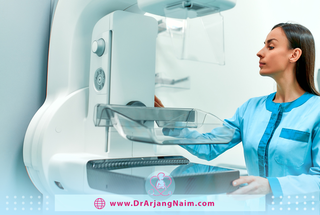
At the testing facility, you will be given a gown and asked to remove jewelry and clothing from the waist up. You stand in front of an X-ray machine explicitly designed for mammography for the procedure itself. A healthcare team member will place one of the breasts on a platform and raise or lower the platform to match your height. The head, arms, and torso are positioned in such a way as to allow an unobstructed view of the breast.
Your breast is gradually pressed against the platform by a clear plastic plate. Pressure is applied for a few seconds to spread the breast tissue. Pressure is not harmful, but it can cause discomfort.
The breast should be compressed to even out the thickness and allow the x-rays to penetrate the breast tissue. Compression holds the breast still to reduce motion blur and minimizes the required radiation dose. You will be asked to stand still and hold your breath during the short X-ray exposure.
After the test
After images are taken of both breasts, you may be asked to wait while the care team reviews the quality of the images. If the views are insufficient for technical reasons, you may have to repeat part of the test. The entire procedure usually takes less than 30 minutes. After that, you can get dressed and resume your normal activities.
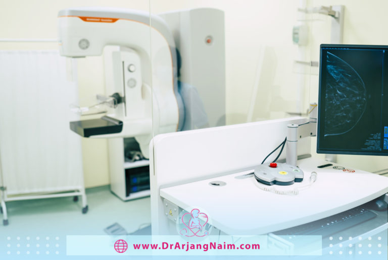
What does the mammography equipment like?
A mammography unit is a box with a tube that produces x-rays. This machine is used exclusively for breast X-ray examinations and has special accessories to limit X-ray exposure to the breast only. It has a device to hold and compress the breast and position it so the technician can capture images at different angles. Breast tomosynthesis is performed using digital mammography units, but not all digital mammography machines are equipped to perform tomosynthesis imaging.
What are the potential hazards of mammograms?
Mammography has potential risks.
Radiation exposure
Mammography requires radiation, and although it is in very low doses and results in low exposure, repeated X-rays have the potential to cause cancer. The benefits of mammography almost outweigh the potential harms of radiation exposure. You should also ensure you are not pregnant because radiation can harm a developing fetus.
False-positive results
A false positive is when a radiologist sees an abnormality that could be positive on an X-ray but is not actually cancer. All mammograms that find abnormalities should be followed up with additional tests to determine if cancer is actually present.
False-positive results are more common among younger women, women with dense breasts, women with a family history of breast cancer, women who have already had a breast biopsy, and women who take estrogen.
A false-positive mammogram can lead to emotional and physical anxiety and distress. The additional tests required to determine the presence or absence of cancer can be expensive, time-consuming, and physically uncomfortable.
False-negative results
A negative result in cancer screening means no abnormality. A false negative result creates the impression that the mammogram is normal despite the presence of breast cancer. This is more common among women with high breast density and younger women because as women age, their breasts become fatter, which reduces the chance of false-negative results.
At what age is mammography performed?
The American Cancer Society recommends annual mammography after the age of 40. The best time to start mammography is at the age of 35, and after the age of 40, it is better to do it once a year and after the age of 50, every two years.
For women from the age of 25, it is better to have other methods than mammography, and if a suspicious case is observed, mammography should be performed, as usually, these people have a history of breast cancer in their relatives.
According to doctors’ advice, people with a history of this disease in their close surroundings should do continuous examinations and even self-exams from 25, the day after the end of the monthly cycle.

Dense breast tissue
Breast tissue consists of milk glands, milk ducts, dense breast tissue, and fat tissue. When seen on mammograms, women with dense breasts have more dense tissue than fatty tissue. In mammography, non-dense breast tissue appears dark and clear. The dense breast tissue appears as a solid white area on a mammogram, making it difficult to see.
What causes dense breast tissue?
It is unclear why some women have dense breast tissue, and others do not. You may be more likely to have dense breasts if you:
- Are younger: Breast tissue becomes less dense with age, although some women may have dense breast tissue at any age.
- Have a lower body mass index: Women with lower body fat have denser breast tissue than obese women.
- Use hormone therapy for menopause: Women who use combination hormone therapy to relieve menopausal signs and symptoms are more likely to have dense breasts.
Why does breast density matter?
Having dense breasts affects in two ways:
- Mammography increases the chance of not detecting breast cancer, as dense breast tissue can mask potential cancer.
- It increases the risk of breast cancer, although doctors aren’t certain why.
What tests are recommended for dense breast tissue screening?
Most medical organizations recommend that women with an average risk of breast cancer start regular mammograms at age 40 and repeat the screening annually. Women with dense breasts but no other risk factors for breast cancer have a higher-than-average risk of developing breast cancer. They may benefit from annual breast cancer screening.
Dense breast tissue makes mammograms more difficult to interpret, as both cancer and dense breast tissue appear white on mammograms. Very dense breasts may increase the risk of not detecting cancer. Despite concerns about detecting cancer in dense breasts, mammography is still an effective screening tool. The most common type of mammography, digital mammography, saves breast images as digital files instead of film, allowing for more detailed analysis.
Complementary tests for breast cancer screening
The patient and the doctor may consider additional or complementary tests based on other risk factors and personal preferences.
3-D mammogram
3-D mammogram uses X-rays to collect multiple images of the breast from multiple angles. The computer synthesizes these images to form a three-dimensional image of the breast. Many mammography centers are converting to 3D mammography as part of standard technology.
Breast MRI
MRI uses magnets to create images of the breast. A breast MRI is recommended for women at high risk of developing breast cancer, such as those with genetic mutations that increase the risk of developing cancer.
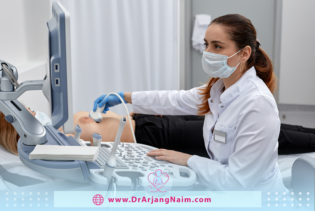
Breast ultrasound
Ultrasound uses sound waves to analyze tissue. Diagnostic ultrasound is usually used to check for areas of concern discovered on a mammogram.
Molecular breast imaging (MBI)
Molecular breast imaging, also known as breast-specific gamma imaging, uses a special camera that records the activity of a radioactive tracer. Cancerous tissue and normal tissue react differently to the detector, which can be seen in the images produced by the gamma camera.
The bottom line
Mammography is the use of low-dose x-rays to examine breast tissue. This test is performed to identify and diagnose breast diseases in women who are faced with abnormalities such as breast pain and nipple depression.
Of course, this test is done periodically to prevent breast cancer in healthy women over 40-45 years old. This test helps detect possible cancerous or benign tumors and breast tissue cysts before they are palpable.
Unfortunately, many people in society are unaware of the benefits of performing physical examinations and periodic mammography imaging to detect breast cancer early.
Dr. Arjang Naim, MD, advises that if you have a history of breast cancer in your family or if you have been placed in a high-risk group by your doctor regarding the risk of this cancer, take periodic mammography imaging seriously.
Additional questions
- How does breast cancer start?
Breast cancer often begins in the cells of the milk-producing ducts. It may also start in a glandular tissue called a lobule or in other cells or tissues of the breast.
- What are the four types of breast cancer?
Types of breast cancer include:
- Ductal carcinoma in situ
- Invasive ductal carcinoma
- Inflammatory breast cancer
- Metastatic breast cancer.
- What vitamin helps dense breast tissue?
Vitamin D may play a role in breast density and breast cancer. Vitamin D reduces proliferation and induces differentiation and apoptosis in breast cells in culture.
- What are the seven warning signs of breast cancer?
- Breast or nipple pain
- Nipple retraction
- Swelling of all or part of the breast
- Dimpling or skin irritation
- Scaliness, redness, or thickening of the nipple or breast skin
- Nipple discharge
- Swollen lymph nodes around the collarbone or under the arm
- How long will a mammogram take?
The entire mammography procedure takes about 30 minutes. Each breast is compressed for only 20 to 30 seconds. While compression can be uncomfortable, it causes the breast tissue to expand and flatten.
References:
https://www.mayoclinic.org/diseases-conditions/fibromyalgia/symptoms-causes/syc-20354780
https://www.healthline.com/health/fibromyalgia