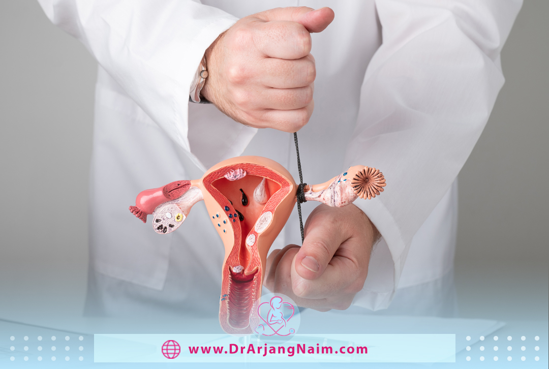
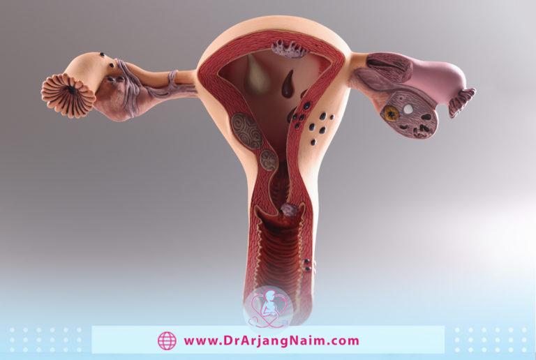
The fallopian tubes are an important passageway for the egg and sperm to meet and for the fertilized egg (embryo) to reach the uterus. Fallopian tube health affects fertility. Damaged or blocked tubes can make it difficult for individuals and couples to conceive.
The fallopian tubes are two thin tubes on each side of the uterus that help guide the mature egg from the ovaries to the uterus. A blocked fallopian tube, also known as tubal factor infertility, is when a blockage, such as adhesion, an ulcer, or infection, prevents the egg from traveling down the tube. It can occur on one or both sides and is the cause of infertility in 30% of infertile people with ovaries.
Anatomy
The fallopian tubes are muscular tubes located in the lower abdomen next to the other reproductive organs. There are two tubes, one on each side, that extend from near the top of the uterus extend laterally and then curve over and around the ovaries.
The open ends of the fallopian tubes are very close to the ovaries but are not directly connected. Instead, the fimbriae of them pull the ovulated eggs into the tubes and toward the uterus.
In an adult, the fallopian tubes are about 10 to 12 centimeters long, although this length can vary from person to person. They generally consist of four parts.
Interstitial section
This part is connected to the inside of the uterus through the wall of the uterus. The interstitial part of the fallopian tube is the proximal part located in the uterus’s muscular wall. Its width is 0.7 mm, its length is 1-2 cm, and it has a slightly winding path from the uterine cavity upwards and outwards.
Isthmus
An isthmus is a narrow section that is about one-third the length of the tube. The part inside the wall (interstitial), which is inside the myometrium of the uterus, is 1 cm long and 0.7 mm wide. The isthmus, which is the lateral continuation of the inner part of the wall, is the round and muscular part of the fallopian tube. Its length is 3 cm, and its width is between 1 and 5 mm.
Ampulla
The ampulla is the main part of the fallopian tube. The ampoule is the widest part of the tube, the maximum diameter of its tube is 1 cm, and its length is 5 cm. It bends over the ovary and is the primary site of fertilization. It contains a large number of ciliated epithelial cells.
Infundibulum
Where the tube expands into a rimmed funnel near the ovary, the margins are known as fimbriae and are sometimes considered the fifth segment. The longest fimbria and the closest fimbria to the ovary is the ovarian fimbria.
Fallopian tubes consist of several layers. The outer layer is a type of membrane known as the serosa. Inside these are layers of muscle known as the myosalpinx. The number of layers depends on the portion of the tube.
Inside the fallopian tubes is a deeply folded mucosal surface. This layer also contains eyelashes. Cilia are hair-like structures. They move to push the egg from the ovary to the uterus. They also help distribute the pipe fluid throughout the pipe.
The cilia of the fallopian tubes are the most numerous at the end of the ovary. They also change during the menstrual cycle. The movement of the cilia increases near the time of ovulation.
Anatomical variations
In rare cases, a collateral fallopian tube can form during development, which can affect fertility. This extra tube usually has an end that is close to the ovary but does not extend into the uterus. Therefore, if the egg is removed by the lateral fallopian tube, it cannot be fertilized and implanted.
This anatomical variation is rare but not unknown, affecting 5-6% of women in some studies. Other changes include additional openings, closed pouches, and fimbria functional changes. There are also cases where one or both fallopian tubes do not develop.

Fallopian tube blockage symptoms
Blocked tubes rarely cause symptoms. The first symptom of blocked tubes is often infertility. If a woman doesn’t get pregnant after a year of trying, a doctor will order a specialized x-ray to check fallopian tubes along with other basic fertility tests.
A specific type of fallopian tube blockage called hydrosalpinx may cause lower abdominal pain and abnormal vaginal discharge, but not all women have these symptoms. It is when a blockage causes the tube to dilate and fill with fluid. This fluid blocks the egg and sperm and prevents fertilization and pregnancy.
However, some causes of blocked fallopian tubes can have their own symptoms. For example, pelvic inflammatory and endometriosis disease (PID) may cause painful periods and painful intercourse.
Symptoms that could indicate pelvic infection include:
- Fever over 101
- Nausea and vomiting
- Severe lower abdominal or pelvic pain
- General pelvic pain
- Pain during sexual intercourse
- Foul-smelling vaginal discharge
What is the role of fallopian tubes?
Fallopian tubes play an important role in pregnancy. Its roles include:
- A holding place for your egg: Each month, one of the ovaries releases a mature egg as part of the menstrual cycle. Finger-like structures at the end of the fallopian tube, called fimbriae, guide the egg into the tube, where it awaits fertilization.
- The site where fertilization happens: If a partner ejaculates during intercourse, his sperm travels through the vagina, cervix, uterus, and finally into the fallopian tubes. When the egg and sperm meet, fertilization occurs in the fallopian tubes.
- An active passageway that moves a fertilized egg to the uterus: A fertilized egg (embryo) travels through the tubes until it reaches the uterus, where it can develop into an embryo. The fallopian tube consists of powerful muscles that move the embryo along.
What makes up the fallopian tubes?
They consist of a thin mucous membrane and layers of muscle.
Mucous membrane
A delicate lining in the fallopian tubes secretes fluids that maintain an environment in which fertilization can occur, and the embryo can develop. Small hair-like structures in the lining (cilia) move in the fallopian tubes, moving eggs, sperm, and the embryo (if fertilization occurs).
Muscular layers
The muscular wall of the fallopian tube has various layers. The outermost layer is mainly smooth and long muscle fibers. The innermost layer consists of circular fibers. These muscles contract together to move the egg, sperm, or embryo through the fallopian tubes with the help of cilia.
Associated conditions
Some medical conditions are associated with fallopian tubes. These conditions can disrupt fertility and, in some cases, can be life-threatening.
Ectopic pregnancy
An ectopic pregnancy is a condition that is mostly associated with the fallopian tubes. It happens when there is a delay in the transfer of the fertilized egg to the uterus. In such cases, the fertilized egg may implant and cause an ectopic pregnancy inside the tube, and there is also a risk of ectopic pregnancy in the accessory tube. An ectopic pregnancy can be very dangerous and can lead to rupture and even death.
Salpingitis
Salpingitis, which is an inflammatory disease, has two types. This disease leads to the thickening of the tubes.
Salpingitis isthmica nodosa
It includes the formation of nodules inside the narrow part of the tubes. These nodules make it more difficult for eggs to pass through the tubes and increase the risk of ectopic pregnancy.
Non-nodular salpingitis
Non-nodular salpingitis is usually caused by an infection, such as those associated with pelvic inflammatory disease. Acute or chronic salpingitis can also cause blocked tubes and scarring.

Tubal infertility
Tubal infertility is a general term that describes when a person is unable to conceive due to problems with the fallopian tubes. It may be for various reasons, from congenital abnormalities to infectious complications.
One of the most common causes of tubal factor infertility is chlamydia complications.
Tubal torsion
Tubal torsion, or adnexal torsion, occurs when a fallopian tube becomes twisted, possibly affecting its blood flow. Although this usually occurs alongside ovarian torsion, it can also occur on its own. If left untreated, tubal torsion can affect fertility.
Hydrosalpinx
Hydrosalpinx describes when one or both fallopian tubes become swollen and filled with fluid. It could be the result of an infection. It can also be caused by the blockage of one or both ends of the fallopian tube.
Cancer
Primary fallopian tube cancer is very rare, but it can occur. Less than 1% of women’s cancers are thought to originate in the fallopian tubes. When cancer occurs in the fallopian tubes, it is most likely the result of metastasis from another site, such as ovarian cancer, uterine cancer, or cervical cancer.
Tests
The problems can be diagnosed with several different tests.
Hysterosalpingogram
A hysterosalpingogram is a special type of X-ray used to examine the fallopian tubes. During this procedure, dye is injected through the cervix. This dye flows through the uterus and into the fallopian tubes. An X-ray is then taken off the dye-filled organs to check for any blockages or problems.
Ideally, a hysterosalpingogram will show that fluid can easily flow through the tubes. If not, there may be problems with fertility.

Laparoscopy
It is a type of surgery that can be used to examine reproductive organs. Small incisions are made, and a camera is placed in the abdomen.
Laparoscopy is often referred to as minimally invasive surgery. It has the advantage that if abnormalities are found during the procedure, the doctor may be able to treat them immediately.
Salpingoscopy
Salpingoscopy involves inserting a rigid or flexible scope into the fallopian tubes. This allows the doctor to see inside the tubes and check for narrowing or blockages. It can be performed during a laparoscopic procedure.
A salpingoscopy also allows the healthcare provider to see how fluid is moving in the tubes.
The bottom line
The fallopian tubes bridge the important work that the ovaries and uterus do. This is why conditions that negatively affect the tubes also negatively affect fertility. Taking steps to prevent infection is the best way to keep your tubes healthy. If your fallopian tubes are damaged or removed, you may still be able to get pregnant through IVF.
Additional questions
- What is the treatment for uterus adhesion?
Intrauterine adhesions are fibrous tissue bands forming in the endometrial cavity, often in response to a uterine procedure. These types of adhesions are often associated with infertility and menstrual abnormalities. Intrauterine adhesions are usually treated by hysteroscopic removal followed by mechanical or hormonal treatments.
- What is Chlamydia?
It is a sexually transmitted disease that can cause infection in men and women and can also cause permanent damage to the female reproductive system, resulting in infertility.
- What are the early warning signs of ovarian cancer?
- Bloating
- Pelvic or abdominal pain
- Trouble eating or feeling full quickly
- Urinary symptoms such as urgency
- Where do eggs go if fallopian tubes are removed?
After surgery, each ovary continues to release one egg. But now, the path of the egg through the fallopian tube is blocked. When the egg and sperm cannot meet, pregnancy cannot occur, and the body absorbs the egg.
- Is fallopian tube removal painful?
After the surgery, there may be a pain in the abdomen for a few days. If laparoscopy is performed, the abdomen may be swollen, or a change in the bowels may occur for a few days. After laparoscopy, there may be some shoulder or back pain.
References
https://www.verywellfamily.com/all-about-blocked-fallopian-tubes-1959927
https://my.clevelandclinic.org/health/body/23184-fallopian-tubes
https://teachmeanatomy.info/pelvis/female-reproductive-tract/fallopian-tubes/
https://www.verywellhealth.com/fallopian-tubes-anatomy-4777161
https://cancer.ca/en/cancer-information/cancer-types/fallopian-tube/what-is-fallopian-tube-cancer


