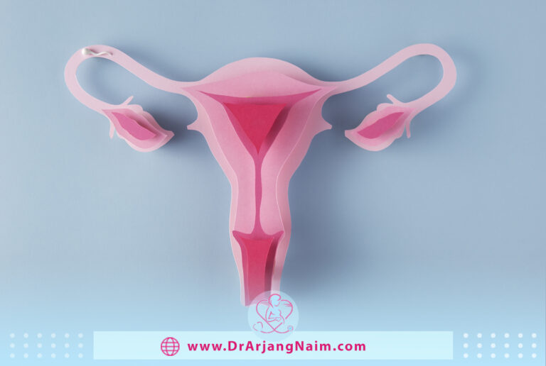
A uterus, also known as the womb, is a hollow, muscular organ present in the female reproductive system of most mammals, including humans. It plays a crucial role in the reproductive process by housing and nourishing a developing fetus during pregnancy. The uterus is where a fertilized egg implants and grows into an embryo and then a fetus.
The shape and size can vary among species, but in humans, it is generally pear-shaped and located in the lower abdominal cavity, between the bladder and the rectum. The upper part of the uterus, the fundus, connects to the fallopian tubes, which transport eggs from the ovaries to the uterus.
During the menstrual cycle, the lining of the uterus, known as the endometrium, thickens in preparation for a potential pregnancy. If pregnancy does not occur, this lining is shed through menstruation. If fertilization occurs and an embryo implants into the uterine lining, the uterus provides a suitable environment for the embryo’s growth and development throughout pregnancy.
What does a uterus do?
It is a vital organ in the reproductive process, and its functions are essential for the continuation of a species through reproduction. The uterus serves several important functions in the female reproductive system, particularly during conception, pregnancy, and childbirth.
Implantation and pregnancy
After fertilization, when a sperm cell successfully merges with an egg cell in the fallopian tubes, the resulting fertilized egg (zygote) travels down to the uterus. It provides a suitable environment for the zygote to implant into the thickened uterine lining (endometrium). This marks the beginning of pregnancy.
Nurturing the embryo and fetus
Once the fertilized egg implants, it is crucial in nurturing the developing embryo and fetus. The uterine lining supplies nutrients and oxygen to the growing embryo through tiny blood vessels, allowing it to develop and grow.
Placenta formation
The placenta, an organ that develops during pregnancy, connects the developing fetus to the uterine wall. It acts as a bridge between the mother and the fetus, facilitating the exchange of nutrients, oxygen, and waste products.
Amniotic fluid
The uterus contains the amniotic sac, which surrounds and protects the developing fetus. The amniotic fluid within this sac provides a cushioning effect, preventing physical trauma and maintaining a stable fetal environment.
Labor and childbirth
As pregnancy progresses, the uterus grows and stretches to accommodate the growing fetus. When the fetus reaches full term, the uterus undergoes contractions, a process known as labor. These contractions help push the fetus through the birth canal during childbirth.
Menstrual cycle
Throughout the menstrual cycle, the uterus changes in preparation for pregnancy. If pregnancy does not occur, the uterine lining is shed during menstruation. This shedding of the endometrial tissue is a normal part of the menstrual cycle.
Overall, the uterus plays a vital role in the reproductive process, from facilitating fertilization and implantation to supporting the growth and development of the embryo and fetus. Its ability to change in size, structure, and function is essential for the continuation of a species through successful reproduction.
What does look like?
The uterus is usually the size of an apple, but during pregnancy, it can stretch to the size of a watermelon. Some conditions may cause it to enlarge, such as cancer, fibroids, and polycystic ovary syndrome. It consists of three distinct tissue layers:
- Perimetrium: The outer layer of tissue made of epithelial cells
- Myometrium: The middle layer is made of smooth muscle tissue
- Endometrium: The inner lining that develops during a month and falls off if there is no pregnancy
Four main parts
The four main parts include:
- Fundus: The broad curved area at the top and widest part of the organ that connects to the fallopian tubes
- Corpus: The main part of the uterus begins directly at the surface of the fallopian tubes and continues downwards, becoming increasingly narrow.
- Isthmus: The narrow lower part of the uterus
- Cervix: The lowest two inches of the uterus, which is tube-shaped and opens into the vagina.
Positions
The uterus can naturally exist in different positions within the pelvis; these variations are considered normal. The positioning of the uterus can be influenced by factors such as genetics, childbirth, pelvic anatomy, and hormonal changes. Here are some common positions:
- Anteverted: This is the most common position of the uterus. In an anteverted uterus, the body of the uterus is tilted forward and leans toward the bladder.
- Retroverted: In a retroverted uterus, the body of the uterus is tilted backward and leans toward the rectum. This position is also common and considered normal.
- Anteflexed: In an anteflexed uterus, the body of the uterus is bent forward at the cervix. This position is often combined with anteversion.
- Retroflexed: In a retroflexed uterus, the body of the uterus is bent backward at the cervix. This position is often combined with retroversion.
- Midposition: Some uteruses are positioned in a more neutral or midposition, with less tilt forward or backward.
It’s important to note that the positions of the uterus can change over time due to various factors, including pregnancy, childbirth, and hormonal changes. In some cases, certain medical conditions or issues, such as uterine fibroids, endometriosis, or pelvic inflammatory disease, can also impact the position of the uterus.
If you are concerned about the position of your uterus or if you are experiencing symptoms related to the position of your uterus, Dr. Arjang Naim, MD, can help you. Dr. Naim will perform a pelvic examination, imaging tests, or other diagnostic methods to evaluate the position and health of your uterus.
Uterus Conditions
The uterus may be susceptible to any number of health problems. Some diseases are more common.

Endometriosis
An estimated 11 percent of women are affected at birth by endometriosis, a condition in which the lining of the endometrium grows outside the uterus.
Fibroids
Uterine fibroids are non-cancerous tumors that grow in the muscle tissue of the uterus. Fibroids often do not cause symptoms or require treatment.
However, for some people, uterine fibroids lead to heavy or painful periods. These symptoms are usually treated with over-the-counter pain relievers containing ibuprofen or acetaminophen, or hormonal birth control. Surgery such as endometrial ablation, myomectomy, or uterine fibroid embolization may be required in severe cases.
Uterine polyps
Polyps are finger-like growths that stick to the uterine wall. They can range in size from the size of a sesame seed to the size of a golf ball. Many people have polyps without knowing it. Uterine polyps have a low risk of cancer and should be removed by a hysteroscopy procedure. Dilation and curettage (D and C) are sometimes performed to remove and biopsy endometrial polyps.
Uterine prolapse
Uterine prolapse occurs when the uterus falls into the vaginal area and sometimes pushes out of the vagina. This happens when the pelvic muscles and tissues are weak.
Uterine cancer
Two types of cancer can affect the uterus.
How big?
The uterus size can vary among individuals and change over a person’s lifetime due to age, hormonal fluctuations, and pregnancy. On average, the dimensions of a non-pregnant adult uterus are approximately as follows:
- Length: About 7 to 8 centimeters (2.8 to 3.1 inches)
- Width: About 5 to 6 centimeters (2 to 2.4 inches)
- Thickness: About 2 to 3 centimeters (0.8 to 1.2 inches)
However, during pregnancy, the uterus undergoes significant changes in size and shape to accommodate the developing fetus. By the end of a full-term pregnancy, it can expand to hold a fetus that weighs several pounds. After childbirth, the uterus gradually contracts and returns to its pre-pregnancy size over a period of weeks to months.
It’s important to note that individual variations in size exist, and the measurements provided are approximate averages. Genetics, hormonal influences, and overall health can contribute to differences in uterus size among individuals.
Symptoms of uterine conditions
Common symptoms of uterine diseases include:
- Difficulty getting pregnant
- Painful urination (dysuria)
- Problems with your menstrual cycle
- Irregular bleeding
- Pelvic pain
- Irregular vaginal discharge
Types of uterine abnormalities
The two ducts merge to form a uterine cavity during normal fetal development. For some people, these ducts don’t line up properly, resulting in an irregularly shaped . Abnormalities of the uterus are congenital. Some of the most common abnormalities include:
- Bicornuate
- Arcuate
- Septate
- Unicornuate
- Didelphys
Tests that diagnose uterine conditions
There are several tests that healthcare professionals use to diagnose conditions of the uterus. These tests help provide valuable information about the structure and function and any potential abnormalities or conditions. It’s important to consult a healthcare provider to determine which tests are appropriate for your situation. Here are some common tests used for diagnosing uterine conditions.

Pelvic examination
A physical examination of the pelvic area, including the uterus, cervix, and surrounding organs. This can help detect any physical abnormalities or signs of disease.
Transvaginal ultrasound
A type of ultrasound where a small, wand-like device is inserted into the vagina to create images of the uterus and surrounding structures. This test can help visualize the thickness of the uterine lining and identify fibroids, polyps, and other structural abnormalities.
Pelvic ultrasound
A non-invasive imaging technique that uses sound waves to create images of the pelvic organs, including the uterus. This can help diagnose conditions such as fibroids, cysts, and tumors.
Hysteroscopy
A thin, lighted tube (hysteroscope) is inserted through the cervix into the uterus. This allows direct visualization of the uterine cavity and can help diagnose conditions like polyps, fibroids, and uterine adhesions.
Endometrial biopsy
A small tissue sample is taken from the lining of the uterus and examined under a microscope. This can help diagnose endometrial hyperplasia, uterine infections, and uterine cancer.
Pap smear
While primarily used to screen for cervical cancer, abnormal results can sometimes indicate uterine or endometrial issues.
MRI (Magnetic Resonance Imaging)
This imaging technique provides detailed cross-sectional images of the pelvic area, including the uterus and nearby structures. It can help diagnose conditions such as adenomyosis and uterine anomalies.
CT (Computed Tomography) Scan
Similar to MRI, CT scans can provide detailed images of the pelvic area to help diagnose various conditions.

Blood tests
Hormone levels, such as estrogen, progesterone, and thyroid hormones, can be measured to evaluate hormonal imbalances or conditions affecting the uterus.
Laparoscopy
While primarily used to diagnose conditions of the abdominal and pelvic organs, laparoscopy can also provide information about the uterus and surrounding structures.
Dilation and curettage (D&C)
This procedure dilates the cervix, and a small tissue sample is scraped from the uterine lining for examination. It can help diagnose various uterine conditions, including abnormal bleeding.
Remember that the recommended tests depend on your symptoms, medical history, and the suspected condition. Discussing your concerns and symptoms with a healthcare professional who can guide you through the appropriate diagnostic process is essential.
The bottom line
The uterus is a hollow, pear-shaped organ behind the bladder and in front of the rectum. It consists of three main tissues and has four separate parts.
It plays a significant role in menstruation, implantation, pregnancy, and childbirth. While the uterus poses certain health concerns, diagnostic tests and treatment options are available.
Additional questions
- Is the fertilized egg a zygote?
The fertilized egg (zygote) divides repeatedly as it travels down the fallopian tube to the uterus. First, the zygote becomes a solid ball of cells. It then turns into a hollow ball of cells called a blastocyst.
- What is the difference between D&C and D&E?
A D&E is performed during the second trimester and is very similar to a D&C in that it uses vacuum aspiration but requires more surgical tools to remove tissue (such as forceps).
- Is an endometrial biopsy the same as a pap smear?
A Pap test, often done with a pelvic exam, is primarily used to screen for cervical cancer. Sometimes, a Pap test may find abnormal glandular cells caused by uterine cancer. Endometrial biopsy A biopsy removes a small amount of tissue for examination under a microscope.
- How do hormonal fluctuations, specifically those related to estrogen and progesterone, impact the uterine lining and its role in the menstrual cycle?
Hormonal fluctuations driven by estrogen prompt the uterine lining to thicken, creating a nurturing environment for potential pregnancy. Progesterone then takes over, further enhancing the uterine lining’s receptivity for implantation. If pregnancy doesn’t occur, progesterone levels drop, causing the uterine lining to shed, leading to menstruation.
- How does the health of the uterus change during different life stages, from puberty to menopause?
The health of the uterus evolves through various life stages. During puberty, it matures for potential pregnancy and menstruation. In reproductive years, it supports pregnancy, adjusts post-childbirth, and manages menstrual cycles. Perimenopause brings hormonal shifts, impacting menstrual regularity and uterine lining. Menopause marks reduced reproductive function, causing the uterus to shrink and undergo structural changes. The uterus further atrophies in post-menopause.
https://teachmeanatomy.info/pelvis/female-reproductive-tract/uterus/
https://www.news-medical.net/health/What-Does-the-Uterus-Do.aspx
https://www.verywellhealth.com/uterus-location-function-female-anatomy-3157180
https://www.betterhealth.vic.gov.au/health/conditionsandtreatments/retroverted-uterus