
Microdeletions are chromosomal abnormalities that can cause health problems. Some prenatal tests look for Down syndrome and other chromosomal abnormalities, but you may not have heard of small deletions. This type of chromosomal disorder is common, but not all microdeletions cause health problems.
What is microdeletion?
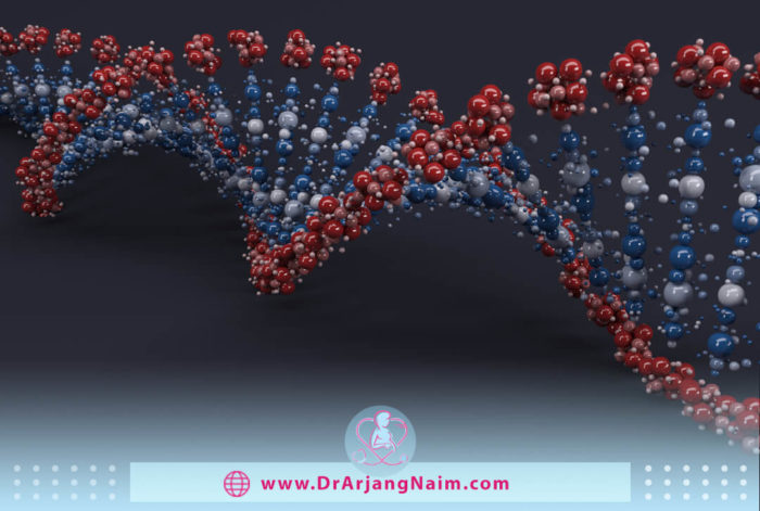
Microdeletion is an abnormality that occurs when a piece of a chromosome is missing. This is exactly what it looks like: micro (tiny), deletion (taken away). Almost all of our cells contain chromosomes with DNA. We get a total of 46 from each parent, 23.
Throughout life, cells proliferate by division, and in the process, the body cuts these strands of DNA to make their use more controllable. But every once in a while, a small number of chromosomes are removed during this process, which leads to the deletion of the micro.
The effect of micro removal on the health and growth of the child depends on its location and size. Some microfibers can cause mental retardation, motor skills problems, or miscarriage, while others do not harm.
What is microdeletion syndrome?

This syndrome is a rare chromosomal disorder. Symptoms may include seizures, moderate to severe learning difficulties, speech delays, behavioral problems, sleep problems, and growth retardation.
The most common microdeletion syndromes are DiGeorge syndrome (22q11. 2), Prader-Willi syndrome, Williams syndrome (7q11. 23), Angelman syndrome (15q11-13), and Wolf-Hirschhorn syndrome (4p16. 3).
DiGeorge syndrome

DiGeorge Syndrome is a disorder that occurs when a small part of chromosome 22 is missing. This removal leads to poor growth of several body systems.
Medical problems commonly associated with 22q11.2 deletion syndrome include heart defects, poor immune function, cleft palate, complications of low blood calcium levels, and stunted growth, along with behavioral and emotional problems.
Signs
The signs and symptoms of DiGeorge Syndrome can vary in type and severity, depending on what systems of the body are affected and how severe the defects are. Some signs and symptoms may appear at birth, but others may not appear until late infancy or early childhood. Signs and symptoms may include a combination of the following:
- Heart murmur and bluish skin
- Frequent infections
- Delayed growth
- A gap in the roof of the mouth (cleft palate)
- Certain facial features, such as low-set ears, an underdeveloped chin, wide-set eyes, or a narrow groove in the upper lip
- Poor muscle tone
- Delayed speech development or nasal-sounding speech
- Learning delays or disabilities
- Behavior problems
- Delayed development
- Breathing problems
- Difficulty feeding, gastrointestinal problems, and failure to gain weight
Treatment
Although there is no cure for DiGeorge syndrome, treatments can usually correct vital problems such as heart failure or cleft palate. Other health and developmental issues, mental health, or behavioral problems can be addressed or monitored as needed.
The number and severity of symptoms associated with 22q11.2 elimination syndrome vary. However, almost all people with this syndrome need to be treated by specialists in various fields.
Prader-Willi syndrome
Prader-Willi syndrome is a rare genetic disorder that leads to several physical, mental, and behavioral problems. One of its main features is the feeling of constant hunger, which usually begins around the age of two. People with the syndrome want to eat constantly because they never feel full. Many of the complications of Prader-Willi syndrome are due to obesity.
Symptoms
Symptoms vary from person to person. They may change slowly over time from childhood to adulthood.
Infants
Signs that may be present from birth include:
- Poor muscle tone
- Distinct facial features
- Poor sucking reflex
- Generally poor responsiveness
- Underdeveloped genitals
Early childhood to adulthood
Other features of Prader-Willi syndrome appear in early childhood and persist throughout life and require careful management. These features may include the following:
- Food craving and weight gain
- Underdeveloped sex organs
- Poor growth and physical development
- Cognitive impairment
- Delayed motor development
- Speech problems
- Behavioral problems
- Sleep disorders
Treatment
Early diagnosis and treatment can improve the quality of life of people with Prader-Willi syndrome. A team of health professionals will likely work together to manage the disease.
This team usually includes a doctor who treats hormonal disorders, behavioral specialists, a nutritionist, a physiotherapist, an occupational therapist, a mental health specialist, a geneticist, and other specialists if needed. Although specific treatments vary depending on the symptoms, most children with Prader-Willi syndrome need the following:
- Good nutrition for infants
- Human growth hormone (HGH) treatment
- Sex hormone treatment
- Weight management
- Treatment of sleep disturbances
- Behavior management
- Mental health care
Williams syndrome
Williams syndrome is a rare neurological and genetic disorder characterized by many symptoms, including unique physical characteristics, growth retardation, cognitive challenges, and cardiovascular abnormalities. It may cause stunted growth in childhood, and most adults with the disease are shorter than average. It can also cause endocrine concerns such as too much calcium in the blood and urine, hypothyroidism, and premature puberty.
Symptoms
Williams syndrome may cause symptoms that vary in severity. Symptoms of Williams syndrome include:
- Chronic ear infections and hearing loss
- Dental abnormalities
- Elevated calcium level in the blood
- Endocrine abnormalities
- Feeding difficulties in infancy
- Scoliosis
- Sleep problems
- Unsteady walk
Treatment
If a person has Williams Syndrome based on physical findings or a positive genetic test result, the health care provider will work with appropriate specialists to explain the diagnosis to the individual and their family, especially a genetic counselor.
Although there is no cure for Williams syndrome, it is important to identify and treat the side effects that may occur. Treatments and interventions should be based on each individual’s unique needs.
Angelman syndrome
Angelman syndrome is a genetic disorder that causes growth retardation, speech and balance problems, mental retardation, and sometimes seizures. People with Angelman syndrome often smile and have a happy personality. Growth retardation, which begins between 6 and 12 months, is usually the first sign of Angelman syndrome. Seizures may start between the ages of 2 and 3.
Symptoms
Signs and symptoms include:
- Frequent smiling and laughter
- Happy, excitable personality
- Sucking or feeding difficulty
- Intellectual disability
- No speech or minimal speech
- Difficulty walking, moving, or balancing well
- Developmental delays
- Trouble going to sleep and staying asleep
Treatment
There is no cure for Angelman syndrome. Current therapy focuses on managing medical issues and growth. A multidisciplinary team of health care professionals will likely work together to manage the child’s condition. Depending on the signs and symptoms, treatment for Angelman syndrome may include the following:
- Physical therapy to help with walking and movement problems
- Anti-seizure medication to control seizures
- Communication and speech therapy
- Behavior therapy
- Medications and sleep training to manage sleep issues
Wolf-Hirschhorn syndrome
Wolf-Hirschhorn is a rare genetic disorder that a child develops by deleting part of chromosome 4. It occurs when cells divide abnormally during reproduction.
Symptoms
The most common symptoms of facial abnormalities are growth retardation, mental retardation, and seizures. Other problems include:
- Bulging, wide-set eyes
- Droopy eyelids and other eye problems
- Cleft lip or palate
- Scoliosis
- Heart and kidney problems
- Failure to thrive
- Downturned mouth
- Low birth weight
- Microcephaly, or an unusually small head
- Underdeveloped muscles
Treatment
There is no cure for Wolf-Hirschhorn syndrome, and each patient is unique, so treatment plans are designed to manage the symptoms.
What causes microdeletions?
Microdeletion syndrome is caused by a chromosomal change in which a small chunk of chromosome 5 is removed in each cell. Deletion in the long arm (q) of the chromosome occurs at the position designated q31.3. The deletion size can vary from a few thousand to several million blocks of DNA (base pairs). The deleted region usually contains at least three genes. The loss of one of these genes, PURA, is thought to lead to most of the hallmarks of the disease.
A protein produced by the PURA gene called Pur-alpha (Purα) plays several roles in cells, including controlling the activity of genes (gene transcription) and helping to copy (replicate) DNA. This protein is especially important for the normal growth of the brain. It promotes the growth and division of nerve cells (neurons) and may play a role in the formation or maturation of myelin.
Loss of a copy of the PURA gene is thought to alter normal brain growth and neuronal dysfunction, leading to growth retardation, hypotension, seizures, and other neurological problems in people with 5q31.3 microdeletion syndrome. Some studies show that the loss of another gene nearby increases the severity of signs and symptoms. It is unclear how the loss of other genes in the deleted region contributes to microdeletion syndrome.
How common are microdeletions?
Unlike other chromosomal abnormalities, which are more common in mothers with certain risk factors (such as those 35 years of age or older with a family history of genetic disorders), microdeletions occur randomly and equally to any baby, regardless of other factors.
By some estimates, almost all of us have some deletion in our chromosomes. For example, 22q11.2 deletion syndrome is rare and occurs in about one in 4,000 people. But this condition may be less diagnosed, meaning that it may occur more often.
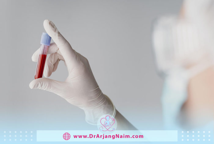
Noninvasive Prenatal Testing (NIPT)
NIPT is a prenatal screening that examines fetal placental DNA in a mother’s blood sample to determine if the mother is at increased risk of giving birth to a child with a genetic disorder. However, screening like NIPT cannot determine if a child has a chromosomal abnormality; there is only a possibility of developing that condition.
NIPT screening results can help the mother and doctor decide on the next steps, including whether a diagnostic test such as chorionic villus sampling (CVS) or amniocentesis should be performed.
These genetic tests analyze the baby’s genetic material collected from amniotic fluid or placenta to say with 100% certainty whether the baby has a chromosomal abnormality. However, CVS and amniocentesis are invasive, which slightly increases the risk of miscarriage.
Because NIPT only contains blood samples, it is safe for both mother and baby. The sample is then sent to a lab, where a technician checks the DNA in the blood for signs of abnormality. After the NIPT results return, the doctor will pair them with the results of the first-trimester ultrasound or nuchal translucency screening to determine if more testing is needed. Depending on the results, your doctor may recommend an amniocentesis or CVS follow-up to confirm the result and check for other problems that NIPT cannot diagnose.
How are microdeletions detected in pregnancy?
NIPT is one of the most widely used tests for pregnant women after nine weeks. This blood test provides information about the health of a growing child.
NIPT (Non-invasive prenatal testing) screens for the most common chromosomal disorders: trisomy 21 (Down syndrome), trisomy 18 (Edwards syndrome), and trisomy 13 (Patao syndrome). An extended NIPT displays five pairs of chromosomes and may detect specific microdeletions. But NIPT is just one of the tests that a doctor can perform to inform the mother about a growing baby and possible health consequences. Although NIPT has a 99% overall detection rate of chromosomal abnormalities, it is not definitive.
The bottom line
In the nucleus of each cell, the DNA molecule is packed into thread-like structures called chromosomes. Each chromosome is made up of DNA tightly wrapped around a histone protein several times to support its structure. While some chromosomal abnormalities are harmless, some are associated with clinical abnormalities. Half of all spontaneous miscarriages are due to chromosomal abnormalities.
One of the chromosomal abnormalities is Microdeletion. It is an abnormality that occurs when a piece of a chromosome is missing. Symptoms and treatments vary depending on the type of chromosome disorder.
Additional questions
1. What are the four types of chromosomal abnormalities?
The four main types of structural chromosomal aberrations are deletion, duplication, translocation, and inversion.
2. What is DNA, and what’s its function?
Deoxyribonucleic acid (DNA) is a molecule that contains biological instructions that make each species unique. DNA is passed from adult organisms to their offspring during reproduction, along with its instructions.
3. What is Down syndrome?
Down syndrome is a genetic disorder that occurs when abnormal cell division results in an additional complete or partial copy of chromosome 21. This extra genetic material causes developmental changes and physical characteristics of Down syndrome. The severity of Down syndrome varies from person to person and can lead to lifelong mental disability and growth retardation. It is the most common genetic chromosomal disorder and the cause of learning disabilities in children. It also commonly causes other medical disorders, including heart and gastrointestinal disorders.
4. What is Chorionic villus sampling (CVS)?
CVS is a prenatal test in which a sample of chorionic villi is removed from the placenta for testing. The sample can be taken through the cervix (trans cervical) or the abdominal wall (transabdominal).
The placenta provides oxygen and nutrients during pregnancy and removes waste products from the baby’s blood. Chorionic villi are the thin protrusions of placental tissue that share the genetic structure of the baby. This test can be done early in the 10th week of pregnancy.
Chorionic villus sampling can show whether the baby has a chromosomal disease such as Down syndrome and other genetic diseases such as cystic fibrosis.
5. What are the four types of genetic disorders?
There are four different types of genetic (inherited) disorders:
1. Chromosome abnormalities
2. Single gene inheritance
3. Multifactorial inheritance
4. Mitochondrial inheritance
References:
https://www.webmd.com/children/wolf-hirschorn-syndrome
https://www.mayoclinic.org/diseases-conditions/angelman-syndrome/diagnosis-treatment/drc-20355627
https://my.clevelandclinic.org/health/diseases/15174-williams-syndrome
https://www.mayoclinic.org/diseases-conditions/digeorge-syndrome/symptoms-causes/syc-20353543
https://www.whattoexpect.com/pregnancy/pregnancy-health/noninvasive-prenatal-testing/
https://www.whattoexpect.com/pregnancy/microdeletion/
https://www.mayoclinic.org/diseases-conditions/down-syndrome/symptoms-causes/syc-20355977
https://www.medicinenet.com/genetic_disease/article.htm